Infertility due to sexually transmitted infections is a problem, but it can be corrected with proper treatment. The disease can occur unnoticed by the woman and the disease is first suspected during an ultrasound scan of the fetus. A pregnant woman can transmit the pathogen to the fetus, if symptoms of the disease appear in the form of disruption of its development or it dies, then this condition is called intrauterine infection.
If a pathogen is transmitted to a sick expectant mother through the general bloodstream, but no clinical symptoms develop, this will be an intrauterine infection. Such infection after the birth of a child may manifest itself as an intensification of the infectious process. In this case, the pregnant woman’s immunity is strong enough to protect the child developing in the uterus.
The international clinic Medica24 performs a full range of tests to detect all infectious diseases. The examination must be completed before pregnancy, but if a woman has missed the favorable time, this does not mean that she cannot be helped. Contact the Infectious Diseases Center by phone.
Why doesn’t a pregnant woman’s immune system protect?
The immune system is weakened during this period, so nature preserves future offspring that are not entirely identical in genetic makeup. Strong immunity would not allow the fertilized egg to attach. For some viruses, embryonic tissue is ideal for reproduction, where all processes are very active and rapid. When they penetrate the pregnant uterus, they can cause lesions, most often in the initial period resulting in miscarriage, and later developmental arrest or birth defects.
Not only the tropism of the pathogen to tissues can lead to damage, but also the release of toxic products and the breakdown of the mother’s cells; it is very important at what period of organ formation the disease began. It is not always possible to identify pathology using ultrasound; in some cases, clarifying tests are necessary to allow a targeted search.
Who should not be vaccinated and when?
Measles vaccination is not recommended for:
- high individual sensitivity to aminoglycoside antibiotics;
- primary immunodeficiency;
- active form of tuberculosis;
- malignant tumors;
- malignant blood pathologies;
- a violent vaccine reaction - with a body temperature above 40°C, local inflammation with a diameter of 8 centimeters or more at the injection site - to the previous administration of the measles vaccine.
It is recommended to delay measles immunization if:
- acute diseases and relapses of chronic pathologies - until the onset of a period of remission or complete recovery;
- pregnancy - due to an alleged risk to the fetus;
- administration of blood products, including immunoglobulins - vaccination is done three months after their use;
- treatment with high doses of corticosteroids - a month after their withdrawal;
- therapy with immunosuppressive drugs - 4-6 months after its completion.
The measles vaccine is made using quail (in Russia) or chicken (abroad) embryos. If there is a violent allergic reaction to the egg white of one or another type of bird, a domestic or imported drug is chosen for vaccination.
Measles vaccine
If there are no clinical symptoms of allergy, but immunoglobulins to bird protein circulate in the blood, measles vaccination is carried out routinely along with desensitizing therapy.
In cases where a woman has an absolute medical exemption from measles vaccination, it is recommended to vaccinate her family members so that they do not serve as sources of infection for the pregnant woman, and subsequently for the newborn.
Disease during different periods of pregnancy
Any infectious disease during pregnancy is dangerous, because an increase in temperature just above 37.7°C already threatens spontaneous miscarriage or premature birth.
- In the first week after fertilization, intrauterine infection will not allow the fertilized egg to attach to the wall of the uterus, and pregnancy simply will not take place.
- In the first two months, infection will destroy the embryo or develop developmental abnormalities.
- By six months, the formation of organs has already occurred, but they are not fully formed, so an infectious disease can lead to the development of pathological changes in them.
- From the 28th week, the fetus acquires its own immunological protection, responding to the introduction of the agent with changes in tissues, which is also manifested by its death, growth retardation, premature birth or congenital infection.
Is coronavirus dangerous for pregnant women?
Respiratory diseases in pregnant women are generally more severe and cause more complications. Doctors explain this by saying that during pregnancy the immune system weakens in order to protect the fetus from the risks of an accidental attack from the mother’s immune system. The downside of this mechanism: the body becomes more susceptible to colds and infectious diseases.
In general, the risk of contracting a coronavirus infection in pregnant women is the same as in everyone else. They are often asymptomatic, and if they have symptoms, they are mild or moderate. The most common clinical manifestations of the disease are fever, cough and myalgia (muscle aches). In addition, fatigue, diarrhea, shortness of breath, and sore throat are common. Overall, most pregnant women experience COVID-19 in the same way as others.
But the chances of getting seriously ill and dying from COVID-19 are still higher. An international study organized by scientists at the University of Oxford and conducted in 18 countries (including Russia) showed that the risk of severe complications is increased in both mother and child. And according to a comparative review of studies involving 10 thousand pregnant and 128 thousand non-pregnant women, the former have almost twice the risk of death. It is just over 11%.
What are the most common infections during pregnancy?
- Chicken pox without complications is not dangerous for the mother and does not lead to miscarriage, but a third of newborns die if the pregnant woman gets sick a week and a half before giving birth. Chickenpox at the beginning of pregnancy in every twentieth person leads to atrophy or disfiguring changes in the limbs, underdevelopment of the cerebral cortex. If a woman has been in contact with an infectious patient, immunoglobulin is administered.
- Viral hepatitis increases the risk of prematurity; they do not cause developmental abnormalities, but lead to congenital hepatitis.
- Influenza is dangerous for the woman herself, since a decrease in immune defense contributes to the introduction of pneumococcus and the development of severe pneumonia with a fatal outcome. The virus passes through the placenta and a miscarriage or premature birth may occur, but the child does not have any abnormalities. But taking amantadine by a pregnant woman can lead to abnormalities in the development of systems and organs. Vaccinations against influenza and pneumococcus help prevent severe pneumonia.
- Rubella in a pregnant woman is no more severe than in others, but the frequency of miscarriage and death increases many times over; if this does not happen, the infection will result in numerous defects. In the third trimester, a favorable outcome for the child is possible. All young women should be vaccinated before pregnancy.
- Measles before childbirth threatens congenital infection, the rest of the time it leads to spontaneous abortion and prematurity, but developmental anomalies are not typical. However, if a woman has not had measles, she needs to be vaccinated at the stage of pregnancy planning.
- Cytomegalovirus infection allows you to carry a child to term, but with intrauterine infection, death or underdevelopment of the brain is possible. Asymptomatic infection in a pregnant woman leads to neurological pathology with visual and hearing impairment in every tenth newborn.
- Mumps causes complications in women; early in pregnancy it can cause miscarriage, but developmental anomalies are not typical.
- Chlamydia can lead to intrauterine infection, but clinical symptoms will appear at birth in the form of mild damage to the eye mucosa or pneumonia.
- Borreliosis or Lyme disease increases the likelihood of fetal death and miscarriage, but pregnant women tolerate it without any special features.
Prenatal viral infection
Ultrasound machine HM70A
Expert class at an affordable price.
Monocrystal sensors, full-screen display mode, elastography, 3D/4D in a laptop case. Flexible transformation into a stationary scanner with a cart.
A distinction is made between intrauterine infection and intrauterine infection of the fetus and newborn. In these conditions, infection occurs during pregnancy or childbirth, and its source is the mother. Intrauterine infection of the fetus
is characterized only by the fact that an infectious agent enters the fetus’s body, but the fetus does not get sick, which is probably due to the activation of protective mechanisms in the mother-placenta-fetus system.
With intrauterine infection,
as a result of infection, a disease develops with corresponding clinical manifestations and damage to the fetus and newborn. At the same time, intrauterine infection occupies one of the leading places in the structure of causes of perinatal morbidity and mortality of the fetus and newborns. The nature of the clinical manifestations and the severity of the disease depend on the type of pathogen, its activity, the severity of the infection, the routes of infection into the pregnant woman’s body and the severity of her body’s defenses.
With intrauterine infection, which occurs in the first 3 months of pregnancy, true malformations occur, primary placental insufficiency is formed, pregnancy does not develop, and spontaneous miscarriages occur. With the development of intrauterine infection after 3 months of pregnancy, intrauterine growth retardation, secondary placental insufficiency are formed, and signs of infectious damage to the fetus are noted. The most characteristic clinical symptoms of intrauterine infection are: polyhydramnios or oligohydramnios, persistent tachycardia in the fetus, delayed fetal development, impaired respiratory and motor activity.
Immunological tests are used to diagnose intrauterine infection.
to identify specific antibodies to pathogens,
the cultural method (seeding)
, as well as
the polymerase chain reaction (PCR) method
.
For the study, blood and material taken from the vagina and cervical canal are used. In some cases, amniotic fluid obtained by amniocentesis is used for research. Ultrasound
is also widely used for diagnosis . Let us dwell on the most typical viral diseases of the embryo and fetus.
Rubella
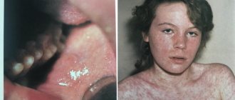
The rubella virus poses perhaps the greatest danger to the embryo and fetus from the point of view of developmental anomalies and significant damage to the fetus. The risk of rubella infection in a pregnant woman is observed in the absence of antibodies to the rubella virus in the mother's blood. If rubella disease occurs in the first 2 months of pregnancy, then the probability of infection of the embryo reaches 80%, and the occurrence of developmental anomalies is possible with a probability of 25%. Infection of an embryo with the rubella virus can lead to its death or leads to the formation of congenital heart defects, deafness, cataracts, microophthalmia, chorioretinitis and microcephaly. Infection of the fetus at a later date may be accompanied by the appearance of typical skin rashes in the newborn, which disappear after some time.
Taking into account the high risk of developmental anomalies during the disease in the first 2-3 months of pregnancy, it must be interrupted. A child who was born to a woman who had rubella during pregnancy is himself a carrier of the virus, and therefore his isolation is necessary. In case of contact of a pregnant woman with a patient with rubella, if she has not had it before, vaccination is necessary, but not earlier than 8-10 weeks of pregnancy, since a live attenuated vaccine is used for this purpose, and a negative effect on the embryo is possible.
Cytomegalovirus (CMV)
Cytomegalovirus (CMV) is one of the most common viruses transmitted to the fetus. When infected with CMV, there is a high probability of non-developing pregnancy, spontaneous termination, premature birth, fetal death, and abnormalities of its development. Detection of CMV in the body of a pregnant woman does not mean that she has an acute disease, since in most cases there is only asymptomatic virus carriage or subclinical chronic infection. In 10-20% of whom specific antibodies to CMV are detected in the blood, an exacerbation of the disease may occur, and the development of intrauterine infection in only 1-2% of these pregnant women. CMV infection occurs for the first time in 1-4% of pregnant women, and accordingly, 40-50% of them have a risk of developing intrauterine infection.
Mortality in congenital CMV infection reaches 20-30%. 90% of surviving children experience late complications such as hearing loss, mental and physical development delays, optic nerve atrophy, dental growth defects, etc. When managing pregnant women with CMV, the specific antiviral drug acyclovir is used only for strict life-saving indications due to the condition of the mother and newborn. It is also possible to use immunomodulators based on recombinant alpha-2 interferon, which is administered in the form of suppositories into the rectum for 10 days.
Herpes simplex virus (HSV)
The causative agent of genital herpes is most often HSV type 2. In 15% of cases, the disease is caused by HSV type 1. In 90% of cases, children become infected during childbirth due to direct contact with infected tissues of the birth canal. About 5% of children become infected during pregnancy. In the remaining 5% of cases, infection occurs after childbirth as a result of contact with others and mainly with the sick mother. The risk of infection during pregnancy depends on the nature of the infection. Thus, the risk of transmitting the virus to a child during a first-time infection with clinical manifestations at the time of birth is up to 50%. For primary infection with asymptomatic course - 40%. If during pregnancy there was a relapse of genital herpes with clinical manifestations, then the risk is 3%, and with a recurrent asymptomatic course - 0.05%. Under the influence of a herpetic infection, damage to the placenta and fetus occurs, which can lead to the formation of malformations, non-developing pregnancy, and its spontaneous termination at various times.
When a child is infected after 32 weeks, the newborn has skin manifestations in the form of herpetic rashes or ulcerations, eye damage - cataracts, microphthalmia, severe disorders of the central nervous system - hydrocephalus, microcephaly, tissue necrosis, calcareous inclusions. In children, severe manifestations of infection such as meningoencephalitis and sepsis may occur. Subsequently, children often experience severe neurological disorders, visual impairment, and developmental delays. A pregnant woman with genital herpes should be informed about the possible risk of transmitting herpes infection to the fetus and about the possibility of a cesarean section. During pregnancy, antiviral drugs are used only in the presence of severe genital herpes. Treatment during pregnancy should be aimed at preventing relapses and antenatal infection of the fetus. For this purpose, immunoglobulin is administered intravenously, immunostimulants and homeopathic remedies are used. When examining pregnant women in the third trimester, you should definitely check for characteristic rashes on the genitals.
The method of delivery depends on the presence or absence of genital lesions at the end of pregnancy, the release of the herpes virus and the time that has passed since the rupture of the membranes. If signs of fresh herpetic lesions of the genital organs are detected shortly before birth, it is advisable to carry out delivery by cesarean section. In the postpartum period, compulsory breastfeeding of newborns is recommended, regardless of the type of herpetic infection in the mother, since breast milk is a source of antiherpetic antibodies, even if the HSV antigen is detected in it.
Viral hepatitis
Viral hepatitis is a serious liver disease. Depending on the type of virus that causes hepatitis, there are the following types: hepatitis A, hepatitis B, hepatitis C, hepatitis D, hepatitis E, hepatitis F and hepatitis G.
Hepatitis A.
The disease is caused by an RNA virus. The infection is transmitted to the mother through the fecal-oral route. Infection of the fetus is rare. Infection of a newborn occurs during breastfeeding while the virus is in the patient’s blood. The incubation period is 15-45 days. In pregnant women, the disease usually occurs in a mild or moderate form. Nausea, vomiting, liver enlargement, jaundice, and pain in the right hypochondrium are noted. Due to the fact that the hepatitis A virus does not penetrate the placenta, it does not lead to malformations in the fetus. Acute viral hepatitis A is cured after a short viremic phase, does not become chronic and does not cause cirrhosis of the liver. Diagnosis of acute hepatitis A is carried out by determining specific antibodies in the blood, which are detected within 2 weeks after infection. Treatment of hepatitis A is carried out according to general therapeutic, symptomatic criteria. In case of contact of a pregnant woman with a patient with hepatitis A, g-globulin is administered for prophylactic purposes.
Hepatitis B
currently represents one of the important health problems, which is associated with an increase in the incidence of the disease and the development of adverse outcomes in the form of the formation of chronic hepatitis, liver cirrhosis and hepatocellular carcinoma. Hepatitis B is caused by a DNA virus. This virus is suspected to be oncogenic. In pregnant women, 1-2 cases of acute hepatitis B are registered per 1000 pregnancies and 5-15 cases of chronic hepatitis B. The source of infection is patients with acute and chronic hepatitis and virus carriers. The virus is transmitted through blood transfusions, blood products, and sexual contact. Infection is also possible through close household contacts (sharing toothbrushes, combs, handkerchiefs) and through the use of poorly treated medical instruments.
In 85-95% of cases, infection of the fetus occurs during childbirth due to contact with blood, infected secretions of the birth canal, or through ingestion of infected secretions. In 2-10% of cases, infection occurs during pregnancy through penetration of the virus through the placenta, especially when placental function is impaired due to fetoplacental insufficiency or placental abruption. In other cases, infection occurs through contaminated breast milk. After childbirth, it is also possible for the child to become infected through contact and household contact from the mother. The severity of the disease in newborns depends on the stage of pregnancy when infection occurred. If the infection occurred in the first or second trimester of pregnancy, the probability of infection is up to 10%. If the infection occurred in the third trimester, then the risk of transmission of infection is 70%. If the HBsAg antigen is detected in the mother’s blood, then the risk of infection of the fetus is 20-40. With the additional presence of the HBeAg antigen, the risk increases to 70-90%.
With hepatitis B, there is an increased incidence of premature births and spontaneous abortions; the number of premature births triples. In most infected children, acute hepatitis B is mild. In the vast majority of cases (90%), children subsequently develop a state of chronic carriage of the virus with the risk of subsequent transmission of infection. There is also a risk of subsequent development of primary liver carcinoma or cirrhosis.
Diagnosis of hepatitis B is based on identifying various antigens and antibodies to the virus in the patient’s blood. If acute hepatitis B develops during pregnancy, therapy consists of supportive treatment (diet, correction of water and electrolyte balance, bed rest). When coagulopathy develops, fresh frozen plasma and cryoprecipitate are transfused. Pregnant women with various forms of hepatitis B should avoid various invasive procedures during pregnancy and childbirth. One should also strive to reduce the duration of the anhydrous interval and labor in general.
The presence of hepatitis B is not an indication for delivery by cesarean section, since it also does not exclude the possibility of infection (contact with infected blood). In the postpartum period, all newborns born to mothers who are carriers of the hepatitis B virus are subject to vaccination. Newborns are also advised to receive the protective immunoglobulin “Hepatotect” in the first 12 hours of life. If vaccinated immediately after birth, breastfeeding should not be avoided. The main method of preventing a child from becoming infected with viral hepatitis B is to screen pregnant women three times for the presence of HBsAg. If there is a risk of infection with the hepatitis B virus in a pregnant woman, it is advisable to vaccinate the patient 3 times with a recombinant vaccine without risk to the child and mother.
Hepatitis C
characterized by a tendency to develop a chronic process, limited clinical symptoms and a poor response to antiviral therapy. Subsequently, the likelihood of developing hepatocellular carcinoma is high.
The causative agent of hepatitis C is an RNA virus. Sources of infection are patients with chronic and acute forms of hepatitis C, as well as latent carriers of the virus. The virus is transmitted through transfusion of infected blood or its components. Contact-household and sexual routes of infection are quite rare. The main route of infection in children is vertical transmission from the mother. The incubation period averages 7-8 weeks. The disease is divided into three phases: acute, latent and reactivation phase. The acute phase in most cases proceeds without clinical manifestations and in approximately 60-85% of cases turns into a chronic form of hepatitis with the risk of developing liver cirrhosis and hepatocellular carcinoma.
Acute hepatitis C, both latent and clinically manifested, in 30-50% of cases can result in recovery with complete elimination of HCV. However, in most cases it is replaced by a latent phase. During the latent phase, infected individuals consider themselves healthy and do not make any complaints. The reactivation phase corresponds to the onset of new clinical manifestations of hepatitis C with the subsequent development of chronic hepatitis, liver cirrhosis and hepatocellular carcinoma.
There is currently no vaccine for hepatitis C. All pregnant women undergo mandatory screening for hepatitis C three times during pregnancy. Despite the fact that vertical transmission of the virus to the fetus is possible, hepatitis C is not a contraindication to pregnancy. The risk of fetal infection with hepatitis C does not depend on the time of infection of the mother and is about 6%. Transmission of the virus is possible both during pregnancy and during childbirth.
There is no consensus on the optimal method of delivery for pregnant women with hepatitis C. Some experts believe that cesarean section reduces the risk of infection of the fetus, while others deny this. Premature rupture of the membranes and a long anhydrous interval increases the risk of transmission of infection.
The hepatitis C virus is also found in breast milk, and in this regard, there is also no consensus on the safety of breastfeeding. All children born to mothers with hepatitis C will also have antibodies to the virus in their blood during the first 12 months of life. If antibodies persist more than 18 months after birth, this confirms that the child is infected with hepatitis C.
Hepatitis D.
The causative agent of the disease is the hepatitis D virus, which is a defective RNA-containing virus that is able to replicate only with the help of the HBsAg antigen of the hepatitis B virus. The infection is transmitted through transfusion of blood or its components, as well as sexually. Infection of the fetus occurs vertically. Diagnosis of viral hepatitis D is based on the detection of antibodies in blood serum. When infected, a newborn develops chronic hepatitis D with a high risk of liver cirrhosis. For hepatitis D, a pregnant woman should be immunized according to the vaccination schedule as for hepatitis B. Treatment of the disease is carried out as part of general therapeutic measures.
Hepatitis E.
The causative agent of the infection is an RNA virus that spreads through the fecal-oral route and causes acute hepatitis. The virus reaches the fetus through vertical transmission. With hepatitis E, the frequency of spontaneous miscarriages is increased. Diagnosis of the disease is based on direct detection of the virus and determination of specific antibodies. Treatment of acute hepatitis E is carried out according to the general principles of symptomatic therapy.
Hepatitis G.
There is a high infectious risk for the newborn with this disease. In the presence of hepatitis G in pregnant women, 33% have vertical transmission to the fetus and newborn. However, to date, no clinical symptoms of hepatitis have been identified in newborns. The presence of the virus in milk was also NOT detected, however, by analogy with hepatitis C, it is advisable to refrain from breastfeeding the child. The diagnosis is made by detecting the virus using PCR. Treatment and prevention of acute and chronic forms of viral hepatitis G have not yet been fully developed.
Flu
Influenza, which can be severe in pregnant women, can lead to damage to the embryo and fetus. With this disease, spontaneous abortion, fetal death, and abnormalities in its development can occur. As a result of infection, the birth of premature and functionally immature children, as well as children with insufficient body weight, is possible.
The effect of the influenza virus during intrauterine infection is due to the influence of pathogens on the placenta and fetus, as well as severe intoxication, elevated body temperature, and impaired uteroplacental circulation with subsequent development of fetal hypoxia. During influenza outbreaks, pregnant women should be immunized with a multivalent killed vaccine.
Parvovirus infection
The causative agent of the infection is parvovirus B19 (DNA virus, family of parvoviruses, genus of erythroviruses), which causes a systemic disease - erythema infectiosum. The infection is most often transmitted by airborne droplets. Persons who work in children's groups, as well as those who have children under the age of 10, are particularly at risk of infection. Parenteral transmission is also possible through transfusion of blood or its components. If infected during pregnancy, the virus can be transmitted transplacentally.
During pregnancy, the clinical picture is characterized by skin rash, sore throat, arthralgia, arthrosis, transient aplastic anemia, and low-grade fever. Pregnancy does not affect the course of the disease. However, if infected during pregnancy, the likelihood of premature termination of pregnancy and intrauterine infection of the fetus increases. The frequency of transmission of the virus to the fetus during acute infection is 33%. Under the influence of a virus that affects the fetal red blood cells, its hemoglobin level decreases. The resulting severe anemia causes dropsy, cardiovascular decompensation and fetal death. The risk of intrauterine infection in the third trimester decreases, which is associated with the formation of the fetal body's defenses. It is possible that there is a relationship between erythema infectiosum in the first trimester and various fetal eye abnormalities.
Diagnosis is carried out using PCR. Detection of specific antibodies to the virus in the blood confirms the diagnosis. The detection of both IgG and IgM indicates an acute infection; the detection of only IgG confirms a past parvovirus infection. There is no specific treatment for the disease. Intrauterine therapy with red blood cell concentrate is carried out when the hemoglobin level is less than 80 g/l. Before treatment, blood samples are taken to detect parvovirus DNA.
Coxsackievirus infection
With coxsackievirus infection, intrauterine infection in the first trimester of pregnancy rarely occurs. However, if this happens, malformations of the gastrointestinal tract, genitourinary system and central nervous system may form. If infected in late pregnancy, a newborn may experience symptoms such as fever, refusal to eat, vomiting, skin rashes, and convulsions. Some newborns may experience otitis media, nasopharyngitis, and pneumonia.
HIV infection
The causative agent is the human immunodeficiency virus (HIV), an RNA virus. There are two types of HIV - HIV-1 and HIV-2, which lead to the development of acquired immunodeficiency syndrome (AIDS). With HIV-1 infection - in 20-40%, and with HIV-2 infection, the disease develops in 4-10% of those infected. This is a disease associated with severe impairment of T-cell immunity in adults and T- and B-cell immunity in children. The source of infection is AIDS patients and virus carriers. Moreover, the period of virus carriage can be very long (years). The virus is mainly spread through sexual contact, as well as through transfusion of blood and its components. The fetus can become infected due to penetration of the virus through the placenta, as well as during and after childbirth through infected milk and through close household contact between mother and newborn.
The course of HIV infection during pregnancy can be aggravated due to the natural weakening of the pregnant woman’s immune defense. The most common and severe complications of HIV infection during pregnancy are: genital candidiasis; cervical neoplasia; premature birth; premature rupture of membranes; delayed fetal development; Chorioamnionitis. The most dangerous complication of pregnancy is infection of the fetus with HIV infection, which is observed in 30-60% of cases, regardless of the presence of symptoms of the disease in the mother. HIV infection can occur during pregnancy, childbirth and the postpartum period. In this case, there are 3 possible ways of transferring the virus to the fetus.
Infection can be transmitted to the fetus through the placenta if it is damaged due to placental insufficiency or placental abruption. Infection is also possible during primary infection of the placenta and accumulation of the virus in Hofbauer cells, followed by the virus multiplying and passing to the fetus. Infection of the fetus can also occur during childbirth when the mucous membranes of the fetus come into contact with infected blood or secretions of the birth canal. After birth, 15 to 45% of children from HIV-infected mothers become infected. Most of these women are unaware of the infection and mainly infect their children through breastfeeding.
HIV infection of a fetus or newborn leads to the development of immunodeficiency, which differs from that of adults. Before 5 years of age, AIDS develops in 80% of children infected with HIV during pregnancy. The first signs of intrauterine HIV infection are low body weight, malnutrition and various neurological symptoms. Soon after birth, persistent diarrhea, lymphadenopathy, enlarged liver and spleen, fungal infection of the oral cavity, and developmental delay occur. Pneumonia and recurrent infections are common. Symptoms of damage to the central nervous system are associated with diffuse encephalopathy, cerebellar atrophy, microcephaly, and the deposition of intracranial calcifications.
Diagnosis of HIV infection is based on identifying risk factors or clinical symptoms of the disease with confirmation of the diagnosis using serological tests. Serological studies are carried out using enzyme-linked immunosorbent assays in combination with confirmatory tests. Currently, prescribing specific antiviral drugs to pregnant women can reduce the risk of intrauterine infection to 5-10%. Such an antiviral drug for pregnant women is zidovudine, an analogue of HIV nucleosides.
HIV infection in a patient receiving antiviral drugs is not an indication for cesarean section in women, since the risk of fetal infection during cesarean section and natural birth is approximately the same. For HIV-infected women who have not received treatment during pregnancy, caesarean section is currently the treatment of choice. In the case of vaginal delivery, you should adhere to the rules for managing childbirth for any viral infections. To prevent postnatal infection, breastfeeding during HIV infection is contraindicated.
In order to reduce cases of intrauterine infection, all pregnant women are required to be tested for HIV infection three times during pregnancy: upon registration, at 24-28 weeks, and before birth. HIV screening of pregnant patients' sexual partners is also recommended.
Ultrasound machine HM70A
Expert class at an affordable price.
Monocrystal sensors, full-screen display mode, elastography, 3D/4D in a laptop case. Flexible transformation into a stationary scanner with a cart.
What needs to be done?
In case of spontaneous abortion due to a “childhood” disease, subsequent pregnancies are not in danger. But to avoid trouble, it is necessary to undergo an examination. When a developmental pathology is identified, it is necessary to do tests for the presence of antibodies to certain pathogens of infectious diseases; however, it will not be possible to turn back the clock, but it is possible to reduce adverse manifestations in relation to the female body.
With chronic infections, you can also safely carry and give birth to a healthy child, but for this you need to undergo an examination in advance and the entire period of gestation is under the supervision of an infectious disease specialist. Specialists at the international clinic Medica24 are able to correct a bad pregnancy history, sign up for a consultation by phone