Prevention
To prevent complications, the catheter installation site is inspected daily and the sutures are treated with antiseptics.
If blood leaks from their wounds, the bandages are changed without delay. To prevent infection, it is necessary to thoroughly rinse the catheter tubes with saline solution after each manipulation:
- administration of antibiotics;
- administration of nutrient solutions;
- administration of blood components.
After rinsing, a small amount of heparin-containing isotonic sodium chloride solution is injected into the tube.
When installing a catheter for a long time, it is recommended to apply a compress with thrombolytic ointments in the area of the puncture and 3-5 cm above it.
General information about the catheter
When should the catheter be replaced? - In most cases, long-term catheters should be replaced every 1-3 months.
What should you do if your catheter is blocked? — If a catheter blockage or urinary tract infection develops, consult your doctor or nurse, who will decide whether additional treatment or catheter replacement is necessary.
Storing Catheters - Catheters should be stored horizontally in a cool, dark, dry place.
Principle of catheter design
First of all, the medical staff should know how to administer intravenous infusion of medications. But if patients know information about the procedure, they may be less afraid.
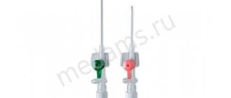
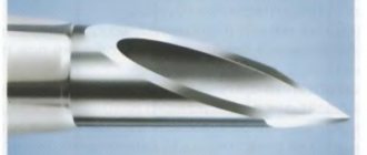
A catheter for intravenous drug administration is a thin, hollow tube. It is inserted into the bloodstream.
This can be done in the arms, neck or head. But inserting catheters into the vessels of the legs is not recommended.
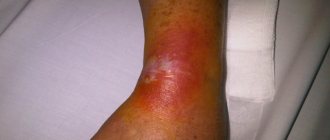
These devices are installed so that there is no need to constantly pierce the veins. After all, this can cause them to become injured and inflamed. Constant damage to their walls leads to thrombus formation.
How to choose the right Foley catheter?
In order to choose the right option, you need to seek recommendations from your doctor. The main criterion is, of course, the duration of use. It can be periodic, temporary or lifelong. Please note:
- What is the purpose of use;
- Gender and age of the patient;
- Catheter size.
Do not neglect the doctor’s advice, as only he can choose the ideal option that will not cause inconvenience to the patient. Here you should take into account both the size of the urethra and the size of the urethra.
Advantages and disadvantages of ports
Manufacturers produce several types of devices designed for intravenous administration of medicinal solutions. According to many, it is preferable to use cannulas equipped with a special port. But it is not always the case. They are necessary if the treatment involves additional infusion of medications.
If this is not required, a regular intravenous catheter can be installed.
- Why do the veins in the arms hurt in the elbow bend?
A photo of such a device makes it possible to see that it is very compact. Devices without additional ports are cheaper. But this is not their only advantage. When using them, there is less chance of contamination. This is due to the fact that the injection element of this system is separated and changed daily.
In intensive care and anesthesiology, preference is given to ported catheters. In all other areas of medicine, it is enough to establish the usual option.
By the way, in pediatrics they can install a catheter with a port for injecting medications even in cases where children do not need to have an IV installed. This is how antibiotics can be injected, replacing injections into the muscle with intravenous administration. This not only increases the effectiveness of treatment, but also facilitates the procedure. It is easier to install a cannula once and almost imperceptibly inject the medicine through the port than to give painful injections several times a day.
Features of catheterization
Regardless of the type and reason for use, all catheters require mandatory fixation. The tube is secured to the skin using medical tape or suture material. Modern models are initially equipped with special clamps, which greatly facilitates the catheterization process. Additionally, it is necessary to set the position of the tube inside the cavity; most often, the instrument has a device that allows you to quickly change its shape after insertion into the hollow organ.
The most widely used system is the Pigtail system - the tip of the catheter made of polyvinyl material looks very similar to a pig's tail, hence the name. During production, this device is placed in a special stylet or conductor, and after installation it is released and, twisting, prevents the tube from falling out of the organ. This type of fixation system is recognized as the safest and easiest to implement.
For a more rigid fixation, a loop is used, which is tightened with a fishing line previously placed in the catheter cavity.
Catheter placement
Catheterization of central veins and peripheral vessels is allowed only in departments of medical institutions. The procedure is performed by a vascular surgeon, anesthesiologist, or interventional radiologist. Before inserting a catheter into a vein, medical workers prepare:
- determine the presence of allergic reactions to administered drugs;
- analyze the degree and speed of blood clotting;
- drugs are prescribed to prevent thrombosis.
If it is planned to place a catheter on a woman, the doctor is obliged to determine the presence or absence of pregnancy.
Regardless of the place where the catheter is placed, the doctor treats the skin with an alcohol solution and then injects it with anesthetics. An incision (for deeply located vessels) or a puncture (for superficially located vessels) is made on the skin above the vessel using the included needle. The tube is installed using a guide. The latter is removed after reaching the required immersion depth.
The final stage of the central venous catheterization procedure is suturing and fixing the device to the skin. A locking cap is placed on the inlet of the catheter. The catheter is then covered with a sterile dressing, on which the current date is stamped. This is necessary to track the period how long you can keep the catheter without reinstallation.
Catheterization of the subclavian vein
The success rate of puncture and catheterization of the subclavian vein reaches 99-100%. The vessel has a fairly large diameter; getting into it is not difficult. Performing puncture and catheterization of the subclavian veins is standard. The patient is placed on the operating table on his back, his head tilted to the side so that the doctor has free access to the injection site.
After local anesthesia, the doctor inserts a needle under the collarbone to a depth of about 4 cm until the costoclavicular ligament is pierced. After this, the needle advance slows down. When the subclavian vein is punctured, the doctor feels another dip in the needle.
- The veins behind the knee hurt
To prevent embolism from occurring during puncture and catheterization of the subclavian vein, the patient should slightly hold back his or her breaths after puncture. The syringe is removed, but the needle remains in place. A conductor is inserted into it, after which the needle is removed, and the catheter is guided onto the remaining guide line using rotational movements. After reaching the required depth, the conductor is removed. The process of puncture and catheterization of the subclavian vein is completed by washing the device tube with saline and fixing it to the skin with silk sutures.
With proper care of the catheter, it can remain in place for up to 2-3 months.
Catheterization of the internal jugular vein
When catheterizing the internal jugular vein (abbreviated as IJV), it is important to be precise and careful with the needle placement. The slightest inaccuracy will lead to rupture of the wall of the carotid artery.
The technique of catheterization of the internal jugular vein involves preliminary anesthesia of the tissues in the area of catheter insertion. As in the previous case, this is done using a 10-gram syringe with anesthetic. The drug is injected into the subcutaneous tissue in the area of the sternocleidomastoid muscles, 5-10 mm outward from the place where the collarbone connects to the sternum. At this point, the jugular vein is located as close to the surface as possible.
As the needle immerses, the doctor should feel two “failures”: when passing through the fascia of the neck and at the moment of penetration through the wall of the vessel. After the second failure, the speed of needle advancement is significantly reduced, and then the steps are repeated to install the guidewire and catheter.
Femoral vein catheterization
Catheterization of the femoral vein begins with the administration of an anesthetic. The doctor places the needle at an angle of 45 degrees to the surface of the skin outward from the place where the pulsation of the femoral artery is felt, that is, on the midline between the pubic fusion and the upper border of the ilium. The needle is inserted to a depth of 2-4 cm until it “falls through”.
Once the needle enters the femoral vessel, it is important to remove the plunger and ensure that it is in a vein and not an artery.
Peripheral vein catheterization
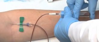
Anesthesiologists and vascular surgeons consider catheterization of peripheral veins to be the simplest procedure, the algorithm of which differs significantly from the insertion of tubes into the central vessels. The procedure does not require local anesthesia. To improve vessel visualization, catheterization of peripheral veins begins with applying a tourniquet above the puncture site. After the contour swells, the doctor inserts a cannula into it at a slight angle. When the needle enters the lumen of a blood tube, dark blood is visible on the needle in the imaging chamber. A catheter for peripheral veins is inserted through the needle. The outer end of the tube is fixed to the skin with an adhesive plaster.
Umbilical vein catheterization
The availability and sufficient size of the umbilical vessels in newborns allows them to be used for measuring hemodynamic parameters, administering nutrients and drugs. The technique of the procedure is somewhat different from others. Before catheterization of the umbilical vein is carried out, it is necessary to prepare the intervention area: treat the field with antiseptics, free the mouth in the umbilical cord stump from blood clots. A catheter is inserted into the lumen of the vein, while simultaneously aspirating the vessel to remove blood clots. With a uniform blood flow, the tube is inserted to the required depth, fixed in the stump and a sterile bandage is applied.
Peripherally implanted central venous catheter (PICC)
A peripherally implantable central venous catheter (PICC) is a minimally invasive device for providing long-term central venous access.
Purpose:
The PIC catheter is intended for short-term or long-term peripheral access to the central venous system for intravenous therapy, administration of contrast agents and allows monitoring of central venous pressure.
Description:
The catheter is implanted in a peripheral vein in the arm and passed to the central (superior vena cava or subclavian) vein. The location of the catheter is on the inner surface of the shoulder. The catheter is used during chemotherapy, long-term drip administration of drugs, long-term parenteral nutrition and frequent blood transfusions. The use of a PIR catheter reduces the risk of complications and provides the possibility of outpatient treatment of patients. Catheter.
Depending on the modification, the PICC catheter consists of one or two tubes up to 60 cm long, made of bioinert polyurethane.
Advantages of the PIC catheter compared to other long-term central venous access devices:
- Increased safety of PIC catheter placement compared to port or tunneled catheter placement.
- Minimal blood loss during PIC catheter placement.
- A single puncture of the vein when installing a PIC catheter and no additional injections throughout the entire course of treatment.
- No need to use special needles (as for ports).
- The universal Luer connector at the end of the catheter makes it easy to connect the infusion system.
- Safe and easy removal of the PIC catheter without surgery or incisions - simply pull on the outside of the catheter to remove it.
- Minimal visibility - the catheter is hidden under clothing (under the sleeve).
OKPD2 - 32.50.13.110: Syringes, needles, catheters, cannulas and similar instruments
Item code KTRU - 32.50.13.110-00005222 : Peripherally inserted central venous catheter
Description of CTRU: A sterile, thin, flexible tube designed to be inserted into a peripheral vein and advanced into a central vein to provide short-term or long-term intravascular access for the administration of medications (antibiotics), chemotherapy agents, nutrients, parenteral solutions, anesthetic fluids, and sometimes for specimen collection. blood, blood pressure and temperature monitoring, and contrast injections; The product is not intended primarily for extracorporeal hemotherapy such as hemodialysis. The device, also known as a peripherally inserted central catheter, typically includes special accessories for insertion/operation of the catheter (eg, Luer connectors, stylet). This is a single use product.
Set contents:
- Central venous catheter.
- Tissue expander (dilator).
- 5 ml syringe (with needle).
- Scalpel.
- Injection caps.
- Holder (catheter clamp).
- Roller clamp.
go to product page
| Shelf life: 5 years | Sterilized by OE |
Sterile, pyrogen-free, non-toxic
Individual packaging: blister . Group packaging: cardboard box.
Manufacturer: China