Content:
- General information
- Composition and release form of the drug
- Methods of using methadone
- The dangers of intravenous methadone injections
- How to restore veins after methadone
Methadone belongs to a group of synthetic opioid drugs with a pronounced analgesic effect.
It has become widespread precisely because of its ability to relieve pain in cases where other pharmacological agents fail. In some European countries and the United States, methadone injections are still used to treat opiate addiction. This technique is based on taking the drug in small doses under the supervision of a doctor, which helps get rid of withdrawal symptoms and gradually overcome cravings for heroin or opium.
General information
Methadone was synthesized in 1937 in Germany. It began to be widely used for analgesic purposes in medicine. Free sale in pharmacies and relatively low cost only contributed to its popularity. But after the discovery of fatal cases of overdose and failure of vital organs after long-term use of the substance, open distribution was stopped, and its production was placed under strict control.
Currently, methadone is produced under various brand names, the main ones being Anadon, Physeptone, and Dolophine. In our country, the drug itself and replacement therapy with its use are considered illegal. Since numerous observations and conclusions of doctors indicate that the compound, due to its strong narcotic effect, causes rapid addiction, irreversible changes in the central nervous system, behavioral abnormalities and physical diseases.
The problem of complex veins
Typically, injections into a vein do not cause problems if you have some experience. But there is a category of people in whom they are not visible or hardly noticeable - the so-called complex veins. The problem is that they are quite inconvenient to inject, which is why complications almost always arise (hematomas or injection of medicine past a vein).
There are several reasons:
- Frequent injections.
- Drugs and the consequences of introducing these substances into a vein.
- Heredity. This is due to the special structure of the venous vessels.
- Excess weight. Due to the fact that a person is fat (including at the injection site), fat blocks the veins.
- Irregular exercise. This is more relevant for women than for men, since they are less likely to do hard physical work.
The reasons may also be temporary. For example, a person takes drugs that increase the tone of the venous wall. He may simply be hypothermic or afraid of the injection.
Such cases are best left to professionals. With the wrong approach, the medicine may not go into the vein, but into the fiber that surrounds it. A large bruise at the injection site is not only an aesthetic problem, but also painful.
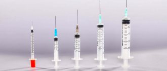
Composition and release form of the drug
Methadone, which is produced industrially at pharmacological enterprises, is purer and safer in terms of additional impurities. The drug is produced in tablet and powder form, as syrup and liquid for injection. The form only affects the method of use, but the active compound is always methadone hydrochloride.
A drug produced by a homemade method carries a greater danger than its pharmacy counterpart. The quality of the chemical is out of the question here, and the purity is highly questionable. Home-grown chemists use unknown reagents and dilute the drug with all sorts of additives to increase the volume and toxic effect. Sometimes even completely different compounds are sold under the guise of methadone. As a result, the home-made drug is tens of times more toxic than the factory-made substance and more often causes poisoning and overdose.
Methods of using methadone
This medication is primarily taken orally or by intravenous injection. It all depends on what pharmacological form the drug has. Drug addicts simply swallow tablets, syrup, or powder or mix them with other psychoactive substances or alcoholic beverages.
The danger of this method lies in irritation of the mucous membranes of the mouth, stomach and intestines. This often causes the formation of foci of inflammation, trophic ulcers, gastritis and even malignant tumors.
For intravenous administration, liquid or powder forms of the drug are used. In the second case, water for injection or distilled water is used for dissolution. Injections allow psychoactive compounds to instantly enter the circulatory system and quickly spread throughout the body.
The duration of action of methadone when administered by injection is much shorter (5-8 minutes) than when administered orally (20-30 minutes). If the substance is used for medicinal analgesic purposes, it begins to act after 40 or 45 minutes and does not retain its effect for 20-30 hours.
A cube of air into a vein: fatal or not?
If you notice that during the injection small air bubbles entered your bloodstream, then you should not immediately panic - there will definitely not be a fatal outcome in such a situation.
Moreover, it makes sense to worry about this only if the intravenous injection was done incorrectly, since the air that gets into the muscle tissue or under the skin almost immediately dissolves in the cells, leaving no consequences, except perhaps short-term discomfort at the injection site. As for intravenous injection, it all depends on the size of the bubble itself. If you let air into a vein just a little, it will immediately dissolve in the cells of the body, as is the case with an intramuscular or subcutaneous injection. That is why the accidental entry of small bubbles into the body will not affect the patient’s health in any way.
The dangers of intravenous methadone injections
If drug addicts inject themselves with the drug, then along with the rapid “arrival” there are the following risks:
- infection in the subcutaneous layer or blood vessels;
- formation of inflammation, abscesses and phlebitis;
- infection with hepatitis or HIV infection;
- formation of necrosis of muscle tissue and subcutaneous tissue;
- overdose or poisoning.
All this happens because among drug addicts they rarely adhere to sterile conditions and allow the use of one syringe for several people. In a hurry or under the influence of withdrawal symptoms, the drug may be injected past the vein. And little thought is given to the correct calculation of the permissible dose and the purity of the substance, when the only desire is to quickly get the required portion.
If during an overdose a person is alone or in the company of inadequate friends, then without timely medical care there is a very high percentage of deaths.
Intravenous injections in Orenburg: what should you know about it?
Intravenous injections
- the most complex type of injection.
Intravenous injections are the most difficult to perform, and a mistake here can lead to dire consequences. Therefore, this procedure should be entrusted to qualified specialists. An intravenous injection can be performed either in a clinic (in a ward or in a treatment room) or at the patient’s home. The last option is especially relevant for pensioners, who sometimes find it difficult to get to a medical facility on their own. Let us note that the prices for this service today can without stretch be called affordable and low.
ATTENTION! All drugs for intravenous administration, as well as their dosage, are prescribed exclusively by the attending physician.
The vein into which the injection is to be made must be clearly visible and contoured. Usually the injection is made into veins in the pits of the elbow, veins of the forearm and hands. Sometimes injections are also given into the veins of the lower extremities, but this is still a rare case.
Let's say the drug needs to be injected into a vein in the fossa of the elbow. How does this happen?
Wherever this procedure is carried out, sanitary and antiseptic rules must be observed.
Disposable syringes and needles must be sterile. For intravenous injections, needles 40 millimeters long are used. The shape of the needles is metal tubes with an oblique cut at one end. The needle is connected to the syringe using a cannula. The needle diameter can be up to 0.8 millimeters. The diameter is determined by the properties of the injected solution. For liquids with good fluidity (this can be a glucose solution, saline solution), thin needles are suitable. For liquids with high viscosity, needles with a larger diameter are used. Note that when installing droppers, a so-called butterfly needle with a length of 2 centimeters and a diameter of 1 millimeter is used.
The nurse performing the procedure should wash their hands and wear disposable gloves.
The patient's skin is pre-treated and disinfected. 70% alcohol works well for this. You also need to prepare cotton or cotton-gauze swabs.
The patient's arm is clamped with a tourniquet, and a cushion is placed under the elbow. The patient can be in either a sitting or lying position.
The patient is asked to unclench and clench his fist so that blood actively flows into the vein.
The very introduction of the solution with the drug occurs quite quickly.
When the drug completely enters the vein, the syringe needle is removed. Then, to avoid the occurrence of hematomas, a swab moistened with alcohol is applied to the injection site.
If the procedure is performed technically correctly, there is practically no pain.
The intravenous method of administering the drug into a vein is justified in several cases:
- when you need to achieve a quick effect. For example, in some emergency situations.
- when the drug is not intended for oral administration (that is, by mouth).
- when its maximum effect can only be achieved when administered into a vein.
As a result of an intravenous injection, the drug in the required quantity enters directly into the blood. And this is the fastest method of achieving maximum concentration. After a short time, the drug reaches the necessary organs and tissues of the body. In addition, the drug administered in this way is eliminated from the body in a very short time.
In medical settings, intravenous injections are given at very reasonable prices. We are happy to take care of your health!
Let us add that the multidisciplinary medical center provides a huge number of services. We work in many areas of medicine: dentistry, gynecology, cardiology, otolaryngology, etc.
How to restore veins after methadone
The process of resuscitating blood vessels after drug injections depends on the degree of their damage and the presence of concomitant diseases. But the main rule for healing is to completely abandon the drug or at least replace intravenous administration with another method of administration.
Only a doctor can assess the condition of the veins and prescribe the correct treatment: narcologist, vascular surgeon, phlebologist. Therefore, if problems are detected, you should not hope that everything will go away over time. Changes can lead to sclerosis and scarring of the walls, thrombophlebitis, and abscesses.
Depending on the complexity of the disease, a patient with affected vessels may need not only conservative, but also surgical treatment. In any case, the medical care plan is comprehensive and, when healing, takes into account all aspects of physical and mental health that manifested themselves against the background of methadone injections.
As additional methods for vein restoration, it is worth considering ointments and gels with antithrombic, anti-inflammatory and bacterial effects, physiotherapeutic procedures, acupuncture, exposure to infrared rays and a magnetic field.
Never take advice from traditional healers or herbalists on how to repair veins after methadone. Some home methods will help relieve painful symptoms and improve the condition of blood vessels. For example, herbal decoctions, compresses and lotions from certain medicinal plants relieve inflammation and reduce suppuration. But before using them, you should definitely consult your doctor and strictly follow his recommendations.
Central venous access in cardiac resuscitation practice
Introduction
Vascular access is called both the line of life and one of the most vulnerable places in patients with various diseases. Despite progress and best practice recommendations, the availability of consistent, secure, and multifunctional access to the vascular bed poses significant challenges to the patient and the healthcare system.
Indications
- Rapid administration of large volumes of fluid
- Administration of vasoactive and irritating drugs to the venous wall, peripheral vein, including parenteral nutrition
- Extracorporeal treatments
- Invasive hemodynamic monitoring
- Transvenous pacing
- Long-term antibacterial therapy
- Transfusion of blood products
- Major surgeries
- Air embolus aspiration
- Inadequate venous access through a peripheral vein
Catheter selection
There are currently many types of catheters available, and their selection should be based on the site of insertion, the reason for use, and the duration of use of the catheter. For example, in anesthesiology and intensive care, the main criteria for choosing a catheter are its length and the number of lumens; catheters with three to five lumens are considered ideal for an ICU patient: this allows several drugs to be administered at once, but the lumens themselves are usually narrow, with high resistance, and therefore less suitable for rapid infusion of drugs during emergency measures (for example, the introduction of drugs that support blood pressure in a strictly calculated dosage, and parallel long-term infusion of antibiotics).
Types of Catheters
- With single/multiple lumens
- Peripherally inserted central catheters (PICCs).
- Tunneled (the catheter is pulled several centimeters under the skin before entering the vessel in order to reduce the risk of infection).
Specialized:
- dialysis catheters
- catheters for long-term continuous invasive monitoring
Choosing a puncture site
There are several access points that can be used for central venous catheterization. The veins themselves can lie deep, pass superficially in the neurovascular bundle, and also next to other anatomical structures. The physician must have a clear understanding of the anatomy of deep and superficial structures to safely perform puncture and catheterization. If available in the intensive care unit, when placing a catheter in the internal jugular, femoral vein, as well as with peripheral approaches, it is useful to use ultrasound. This method helps to visualize the catheterized vessel, confirm the correct placement of the guidewire, and also verify various anatomical variations in location.
The following vessels are most often used for catheterization:
- Internal jugular vein
- Subclavian vein
- Femoral vein
- External jugular vein
- Veins of the upper limb or elbow
Installation principles
The basic preparations and equipment required for central venous catheterization are similar, regardless of placement technique or location. Catheterization should be performed in the selected anatomical area, where aseptic conditions must be ensured. Strict asepsis during catheterization is key to reducing the incidence of infection - use sterile gloves, gown, mask and cap. Correct placement and visualization of anatomical landmarks can reduce the risk of failure and complications. Local anesthesia is used for pain relief.
Procedure for performing the manipulation
Before the procedure, the patient is re-examined for possible allergic reactions to drugs.
- The patient is in a horizontal position.
- The operator and assistant put on a sterile gown, mask, gloves, and cap
- The area in which the manipulation itself will take place is treated with local antiseptics; sterile diapers are used to delimit the required area.
- Infiltration anesthesia with local anesthetics is performed using a sterile needle.
- A puncture needle on a syringe with a solution is inserted in the direction of the vessel at an angle not exceeding 45 degrees, which reduces the subsequent likelihood of excessive bending of the catheter. By tilting the outer end of the needle towards the skin, the anterior wall of the vessel is pierced. Entry into the vessel is confirmed by aspiration of blood into a syringe.
- The needle is tilted even more hollowly, the syringe is removed (or a side channel is used) and a metal conductor is inserted, the tip of which is advanced into the lumen of the vessel 10-15 cm in the central direction. The guidewire usually has a curved J-shaped end, designed to reduce the risk of damage to the vessel wall, as well as to facilitate insertion of the catheter into tortuous vessels.
- The conductor is fixed in the lumen of the vessel, and the needle is removed out. A dilator corresponding to the diameter of the inserted catheter is put on the outer end of the guidewire. The dilator is introduced, moving along the guidewire 2-3 cm into the lumen of the vessel. After removing the dilator, a catheter is put on the outer end of the conductor and, advancing it centrally, the catheter is inserted further into the vascular bed, after which the conductor is removed and further advancement of the catheter is carried out without it.
- Holding the guidewire firmly, the catheter is installed to the required depth, carefully ensuring that the guidewire does not move along with the catheter. When using an introducer, it is placed on the guidewire after removal of the dilator and inserted into the vessel; the next stage is to put a catheter on the outer end of the guidewire and, moving it distally, into the introducer and further into the vessel, then remove the guidewire.
- A syringe is attached to the catheter, and the reverse flow of blood is checked again.
- The catheter is fixed to the skin with a suture, and a bandage with an antiseptic is applied.
Observation
The site of puncture and catheter placement is inspected daily, the condition of the skin is assessed, treatment with antiseptics is carried out, fixing bandages are replaced, the functionality of the catheter is assessed, and rinsing is carried out.
Control
After performing the manipulation, for the purpose of screening control, the patient takes an X-ray of the chest after 1.5-2 hours, with which you can control the position of the catheter and promptly prevent the development of complications after placement if they occur.
Complications
Complications of central venous catheterization develop in 10% of cases and can be divided into mechanical, infectious and thrombotic. The incidence of complications depends on many factors, including the puncture site, patient factors (comorbidities, variant anatomy).
Mechanical complications:
- Arterial puncture
- Hematoma
- Pneumothorax
- Hemothorax
- Bleeding
- Arrhythmia during the procedure
- Cardiac tamponade
- Airway obstruction
- Damage to the thoracic lymphatic duct (lymphorrhea, chylothorax)
- Brachial plexus injury
Infectious complications:
- Local infection
- Bacteremia, sepsis
Thrombotic and embolic complications:
- Vessel thrombosis
- Blood clot formation around the catheter
- Air embolism
- Catheter/guidewire embolism
Conclusion
Central venous catheterization is a vital procedure, however, associated with a number of complications. There are no absolute contraindications to this procedure, as it can be life-saving (as is the case with hemodialysis needed for end-stage renal disease), but serious complications, including death, can occur during or immediately after the procedure. Vascular access plays an important role in the treatment of critically ill cardiac patients. Historically, the preferred method of navigation for central venous catheterization has been the identification of anatomical landmarks, but success depends on the experience and manual skills of the operator, patient comorbidities (eg, coagulopathy, obesity, previous surgery), the environment (eg, resuscitation, mechanical ventilation) and, most importantly, is based on the erroneous assumption that all patients have similar anatomy. As a consequence, the literature reports catheterization failure rates in the range of 7–26%. Currently, numerous international consensus committees are promoting the use of ultrasound guidance in central venous access. As a result, recommendations are being widely implemented in departments of anesthesiology and intensive care, and an increase in the acquisition of necessary equipment and personnel training has been noted. With the advent of cheaper, more portable, higher-quality, high-resolution equipment, and the increased availability of authorized training programs, many barriers to dissemination of the technique have been overcome. It is no longer a new or controversial technique, so everyone involved in emergency cardiac care uses ultrasound navigation when providing central venous access.
Literature:
- Ovcharova, Larisa Germanovna. Causes and consequences of bad habits: textbook / L. G. Ovcharova, L. S. Khoroshilova, Z. A. Kurbatova; Ministry of Education and Science of the Russian Federation, State. higher educational institution prof. education "Kemerovo State. univ." - Kemerovo: Kuzbassvuzizdat, 2006. - 115 p. : table; 20 cm; ISBN 5-8353-0185-5
- Drugs: properties, action, pharmacokinetics, metabolism: textbook / N. V. Veselovskaya [etc.]. — 3rd ed., revised, corrected. and additional - Moscow: Narkonet, 2008. - 262 p. : ill., table; 21 cm; ISBN 978-5-94497-029-9 (translated)
- Sore spot. Vienna. - Nizhny Novgorod: Newspaper world, 2021. - 95 p.; cm.; ISBN 978-5-4423-0242-4